Glaucoma is a major cause of irreversible blindness, impacting millions globally. When medical treatments are insufficient in managing intraocular pressure (IOP), ophthalmic surgical instruments play a vital role in performing precise and effective surgical interventions. Surgeons depend on these specialized tools and advanced techniques to enhance accuracy and safety during intricate procedures. This guide explores the essential instruments and methods used in glaucoma surgery, helping ophthalmic professionals achieve optimal patient outcomes.
Understanding Glaucoma Surgery
Glaucoma surgery aims to reduce intraocular pressure by improving aqueous humor drainage or decreasing fluid production. The two primary types of glaucoma surgery are trabeculectomy and minimally invasive glaucoma surgery (MIGS). Both require high-precision tools designed for microsurgical applications.
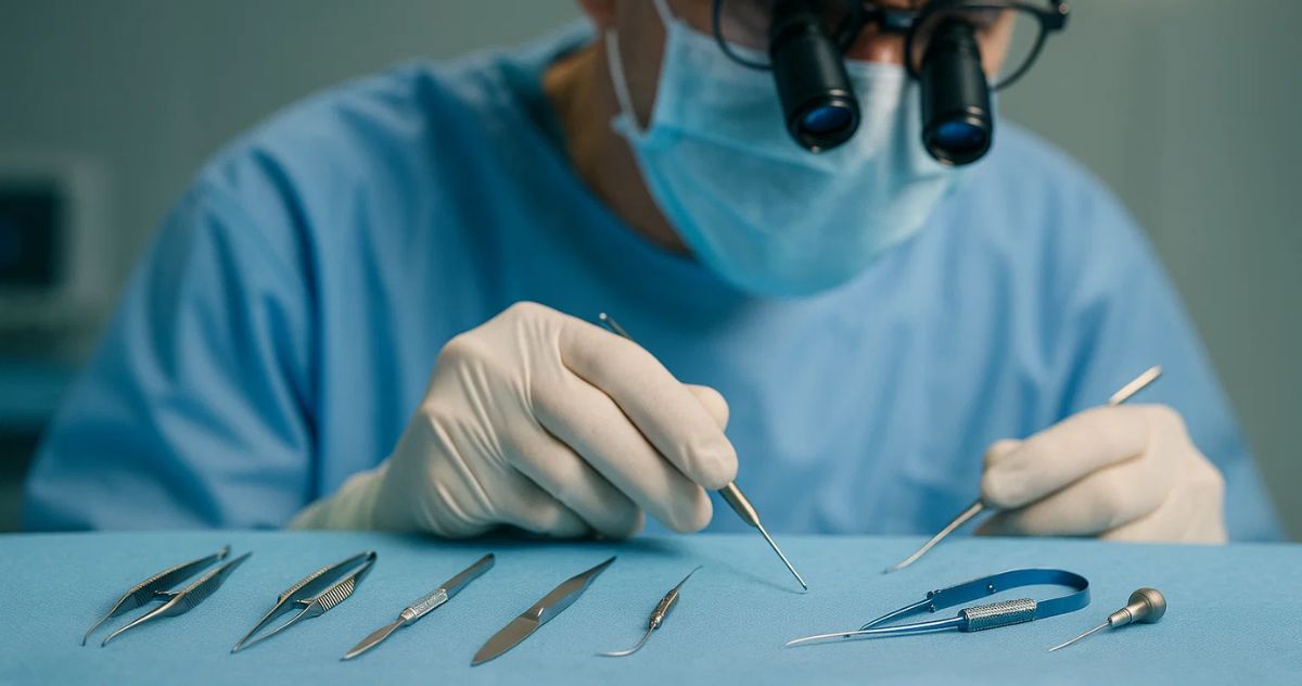
Key Instruments Used in Glaucoma Surgery
1. Microsurgical Forceps
Microsurgical forceps provide precise control when handling delicate eye tissues. They assist in grasping, holding, and manipulating the scleral and conjunctival tissues during surgery. Their fine-tipped design minimizes trauma, allowing for smooth surgical interventions.
2. Trabeculectomy Punch
This instrument creates a drainage hole in the trabecular meshwork to allow aqueous humor outflow, reducing intraocular pressure. The precision of this tool ensures a controlled excision, crucial for long-term surgical success.
3. Scleral Knife
Used to make incisions in the sclera, this knife is designed for controlled penetration, ensuring minimal disruption to surrounding tissues. Different blade angles and sizes are available depending on the surgeon’s preference and the type of glaucoma surgery.
4. Keratome Blade
A keratome blade is utilized in both traditional and minimally invasive glaucoma surgeries to create precise, clean incisions. These blades help in accessing the anterior chamber without causing unnecessary damage.
5. Irrigation/Aspiration Handpiece
This instrument is crucial for maintaining the surgical field by removing excess fluid and debris while ensuring adequate hydration of ocular structures. Proper irrigation and aspiration enhance visibility and reduce postoperative complications.
6. Goniotomy Knife
Used in minimally invasive glaucoma surgeries, this knife facilitates the removal or incision of the trabecular meshwork, improving aqueous humor drainage. The curved or angled design allows access to the drainage system with minimal ocular disturbance.
7. Micro Scissors
Micro scissors allow precise cutting of tissues during trabeculectomy or shunt implantation. These scissors provide enhanced control and ensure that surrounding structures remain undamaged during delicate procedures.
8. Speculum
A speculum holds the eyelids open during surgery, providing an unobstructed view of the surgical site. Adjustable designs enhance comfort for both the patient and the surgeon.
9. Needle Holders
Needle holders are used to secure fine sutures during wound closure. They provide excellent grip and control, ensuring a tight and secure closure to prevent postoperative leaks.
10. Cautery Device
A cautery device is used to control bleeding by coagulating small blood vessels. This helps maintain a clear surgical field and reduces complications associated with excessive bleeding.
Techniques in Glaucoma Surgery
Trabeculectomy: The Gold Standard
Trabeculectomy remains the most commonly performed glaucoma surgery. It involves creating a small drainage flap to allow aqueous humor to bypass the trabecular meshwork, reducing intraocular pressure. The procedure follows these steps:
- Conjunctival Flap Creation: A scleral knife or micro scissors is used to create a conjunctival flap.
- Scleral Flap Dissection: A scleral knife helps dissect a partial-thickness scleral flap.
- Trabeculectomy Punch Use: This instrument excises a portion of the trabecular meshwork, creating an outflow pathway.
- Suturing: Needle holders assist in securing the scleral and conjunctival flaps.
- Application of Antifibrotic Agents: These prevent excessive scarring, ensuring long-term patency of the drainage site.
Minimally Invasive Glaucoma Surgery (MIGS)
MIGS techniques have gained popularity due to their lower risk profile and faster recovery times. They include:
- Trabecular Micro-Bypass Stents: Small stents inserted into the trabecular meshwork to enhance outflow.
- Goniotomy and Ab Interno Trabeculectomy: Procedures using a goniotomy knife or micro scissors to remove obstructions in the drainage pathway.
- Suprachoroidal and Subconjunctival Devices: Implantable shunts that create alternative drainage routes.
These techniques require specialized microsurgical tools to ensure accurate implantation and minimal tissue disruption.
Postoperative Considerations
Instrument Sterilization and Maintenance
Proper sterilization is critical to preventing infections and ensuring instrument longevity. Autoclaving and chemical sterilants are commonly used to maintain sterility.
Suture Management
Fine sutures must be handled delicately using needle holders. Surgeons often adjust suture tension postoperatively to optimize aqueous humor outflow.
Patient Monitoring and Recovery
After surgery, close monitoring of intraocular pressure and wound healing is essential. Patients require careful follow-up to prevent complications such as infection, hypotony, or scarring.
Advancements in Glaucoma Surgery
With the evolution of surgical techniques, modern tools have significantly improved patient outcomes. High-quality surgical tools, developed with precision and ergonomic designs, enhance the efficiency and safety of these procedures. Surgeons continually adopt innovative technologies to refine their skills and optimize results.
Conclusion
Glaucoma surgery requires expertise, precision, and the right instruments for successful outcomes. From traditional trabeculectomy to minimally invasive methods, each procedure benefits from specialized tools designed to enhance accuracy and safety. As technology advances, ENT surgery instruments and other high-precision tools will continue to play a critical role in improving the effectiveness of glaucoma treatments, ensuring better visual outcomes for patients worldwide.