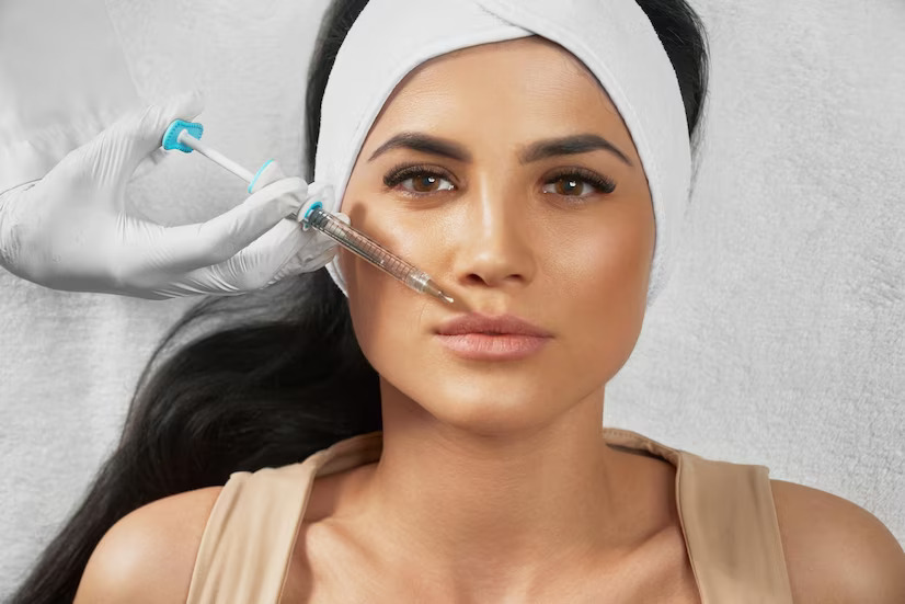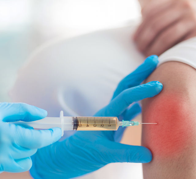
Understanding knee health options is vital for maintaining long-term physical mobility and active living. Many individuals experiencing joint discomfort actively seek modern methods to support the body’s inner healing mechanisms. Exploring targeted biological approaches, such as PRP injections for knees in Abu Dhabi, presents a sophisticated pathway focused on enhancing joint environments and encouraging natural cellular activity.
-
Maintaining joint health requires exploring advanced options for long-term mobility.
-
Individuals look for natural pathways to support cellular recovery and tissue environments.
-
The focus remains on modern biological approaches that work with the body’s natural systems.
Understanding Platelet-Rich Plasma Therapy
Platelet-Rich Plasma, commonly referred to as PRP, is an autologous blood product, meaning it is derived entirely from the individual’s own body. The process involves taking a small sample of blood and placing it into a specialized centrifuge machine that spins at high speeds. This rapid spinning separates the blood into distinct layers, isolating and concentrating the platelets within the plasma portion. The resulting solution contains a significantly higher concentration of platelets and essential bioproteins than what is normally found in whole blood.
-
PRP utilizes autologous blood components directly gathered from the individual.
-
Centrifugation separates and concentrates platelets within the liquid plasma layer.
-
The process creates a rich concentration of natural proteins and signaling cells.
The primary function of platelets extends far beyond their well-known role in basic blood clotting. These specialized cells serve as natural storage units for a multitude of biological signaling molecules known as growth factors. When these platelets are concentrated and introduced into a specific area of the body, they release these proteins to initiate a cascade of natural cellular activities. This targeted concentration serves as a biological signal to alert the surrounding tissues that a localized healing response is required.
-
Platelets house a massive supply of signaling molecules called growth factors.
-
Introducing concentrated platelets initiates a powerful cascade of cellular activities.
-
The solution acts as a targeted signal to prompt localized biological activity.
The Biological Mechanism: Does PRP Repair Knee Tissue?
The question of whether platelet-rich plasma repairs tissue requires a close look at the microscopic cellular processes within the joint. PRP operates by delivering a powerhouse of growth factors directly to the damaged or degenerated areas of the knee. Key proteins inside the solution, such as Transforming Growth Factor-Beta and Platelet-Derived Growth Factor (PDGF), act as biological keys. These keys unlock cellular receptors, encouraging local cells to multiply and begin the intricate process of building new cellular foundations.
-
Growth factors serve as biological keys to activate specific cellular receptors.
-
Essential proteins stimulate local joint cells to proliferate and multiply naturally.
-
The process sets off a microscopic chain reaction designed to build cellular foundations.
Another critical phase of this biological mechanism involves chemotaxis, which is the scientific term for recruiting healing cells to a specific site. The high concentration of signaling proteins released by the platelets acts as a chemical beacon, drawing circulating stem cells and primary repair cells toward the affected knee joint. Once these cells arrive at the destination, they are stimulated to differentiate into the specific types of cells needed for the surrounding environment, assisting the structural network of the joint.
-
Chemotaxis functions as a chemical beacon to recruit native repair cells.
-
Circulating cells are drawn directly to the areas requiring structural support.
-
Recruited cells are guided to adapt to the specific needs of the joint environment.
Furthermore, the introduction of these concentrated proteins supports the process of angiogenesis, which refers to the formation of new, healthy microscopic blood vessels. Areas inside the knee joint, particularly articular cartilage, are naturally avascular, meaning they lack a direct, robust blood supply. By encouraging the development of microvascular networks in the surrounding soft tissues, the joint benefits from an enhanced delivery of essential nutrients, oxygen, and systemic support units necessary for tissue maintenance.
-
Angiogenesis promotes the development of new microscopic blood vessel networks.
-
Improved microvasculature addresses the naturally low blood supply in joint structures.
-
Enhanced nutrient and oxygen delivery provides essential support for ongoing tissue maintenance.
Impacts on Cartilage, Ligaments, and Tendons
Articular cartilage is the smooth, slippery tissue covering the ends of the bones where they meet to form the knee joint. In degenerative joint scenarios, this cartilage experiences progressive thinning and structural breakdown. The growth factors present in a PRP solution interact directly with chondrocytes, which are the specialized cells responsible for producing and maintaining the cartilage matrix. This interaction stimulates the synthesis of Type II collagen and proteoglycans, the fundamental building blocks required to preserve the structural integrity of cartilage surfaces.
-
Specialized cartilage cells are stimulated to synthesize vital structural proteins.
-
The production of Type II collagen helps support the framework of cartilage surfaces.
-
Enhancing these building blocks works to preserve the smooth integrity of the joint.
In addition to cartilage support, soft tissue structures such as tendons and ligaments also respond to the signaling proteins found within plasma solutions. The patellar tendon and the stabilizing ligaments of the knee often experience microscopic tearing or chronic wear over time. The introduction of concentrated platelets encourages fibroblasts—the primary cells found in connective tissue—to increase the production of Type I collagen. This cellular acceleration helps reinforce the internal architecture of tendons and ligaments, promoting improved structural resilience.
-
Connective tissue cells are prompted to increase the output of Type I collagen.
-
Increased collagen production helps reinforce the internal architecture of soft tissues.
-
The structural resilience of key stabilizing ligaments and tendons is enhanced over time.
Enhancing the Joint Microenvironment
Beyond the direct cellular stimulation of structural components, the introduction of concentrated plasma significantly alters the biochemical environment inside the knee joint. In chronic joint conditions, the fluid within the knee often contains high levels of catabolic cytokines, which are destructive proteins that accelerate tissue breakdown and cause discomfort. PRP introduces a balanced wave of anti-inflammatory proteins, such as Interleukin-1 Receptor Antagonist (IL-1Ra), which actively work to neutralize these destructive molecules.
-
The solution alters the internal biochemical balance of the knee joint fluid.
-
Anti-inflammatory proteins work to neutralize destructive, catabolic cytokines.
-
Shifting the environment away from a destructive state protects existing joint structures.
By rebalancing the internal environment, the joint transitions from a state of constant structural degradation to a state of equilibrium and preservation. This shift helps protect the remaining healthy tissue from further enzymatic breakdown while reducing localized swelling. A calm, balanced intra-articular environment allows the native joint cells to function optimally, ensuring that the natural maintenance processes can proceed without being disrupted by chronic biochemical stress.
-
Shifting the joint environment creates a state of equilibrium and structural preservation.
-
Reducing enzymatic breakdown protects the remaining healthy tissue layers.
-
A stabilized environment allows native cells to perform daily maintenance tasks efficiently.
Maximizing the Long-Term Wellness Outcomes
To ensure the best possible response to biological joint care, incorporating a structured physical rehabilitation program is highly recommended. While the concentrated proteins work on a cellular level to optimize the joint environment, physical movement provides the mechanical guidance needed to organize new tissue fibers. Targeted, low-impact exercises help distribute joint fluid evenly across the cartilage surfaces, ensuring optimal nutrient absorption and promoting the gradual strengthening of the surrounding supporting muscles.
-
Structured physical rehabilitation provides mechanical guidance to developing tissue fibers.
-
Low-impact movement ensures even distribution of protective joint fluid.
-
Strengthening surrounding muscle groups reduces overall physical stress on the knee joint.
Individual lifestyle factors also play a substantial role in how effectively the body utilizes the introduced growth factors. Maintaining optimal hydration, consuming a balanced diet rich in amino acids and vitamins, and ensuring adequate rest all contribute to a stronger cellular response. By supporting the body’s baseline systemic health, individuals can maximize the efficiency of the biological pathways activated during their joint care sessions.
-
General systemic health directly influences the efficiency of biological cellular signaling.
-
Proper hydration and a nutrient-rich diet provide the raw materials for cellular activity.
-
Adequate rest allows the body to dedicate energy toward internal joint preservation.
Frequently Asked Questions
How does the biological process of tissue support differ from temporary joint options?
Temporary options often focus on masking discomfort by blocking chemical signals or providing artificial lubrication without changing the joint’s underlying biochemical state. In contrast, biological approaches utilize concentrated growth factors to actively alter the intra-articular environment, neutralizing destructive proteins and stimulating native cells to produce essential structural components like collagen.
When do individuals typically begin to notice structural and functional comfort improvements?
Because this approach relies on natural cellular proliferation, recruitment, and matrix synthesis, the timeline is gradual and progressive. Most individuals begin to observe positive shifts in joint comfort and functional mobility after several weeks, as the internal environment stabilizes and the local tissue structures adapt to the ongoing cellular signaling.
What activities should be prioritized following a biological joint care session?
In the days immediately following a session, prioritizing gentle, low-impact movements is highly beneficial for joint health. Activities such as controlled walking or light stationary cycling help circulate joint fluid without placing excessive mechanical stress on the knee, allowing the introduced signaling molecules to distribute evenly across target areas.
How does the condition of the existing joint tissue influence the overall outcome?
The baseline state of the internal knee structures plays a significant role in the overall success of the session. Individuals with mild to moderate structural changes generally possess a higher density of viable native cells and intact cartilage matrix, providing an optimal foundation for growth factors to latch onto, stimulate activity, and preserve joint longevity.